Home » Without Label » Chest Muscles Anatomy - The Ultimate Mass Building Chest Workout / This mri chest (thorax) axial cross sectional anatomy tool is absolutely free to use.
Chest Muscles Anatomy - The Ultimate Mass Building Chest Workout / This mri chest (thorax) axial cross sectional anatomy tool is absolutely free to use.
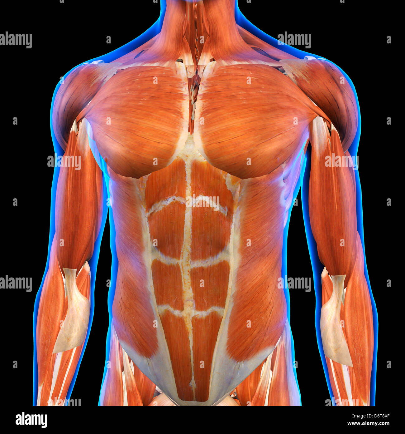
Chest Muscles Anatomy - The Ultimate Mass Building Chest Workout / This mri chest (thorax) axial cross sectional anatomy tool is absolutely free to use.. It contains four muscles that exert a force on the upper limb: Chest muscle anatomy the pectoralis major muscles also known as the pecs are located on the front of the rib cage and form the major muscles of the chest. Learn about each of these muscles, their locations, functional anatomy and exercises for them. Not everyone, however, has the chance to achieve stunning results when working on chest muscles. Let's have a detailed look at each of their types and functions.
The fibers of the pectoralis muscles run like a fan across the. Anatomy chart courtesy of fcit. The beginner as well as advanced players meet with the problem when building a powerful chest. The pectoralis major muscles (also known as the pecs) are located on the front of the rib cage, and form the major muscles of the chest. This mri chest (thorax) axial cross sectional anatomy tool is absolutely free to use.
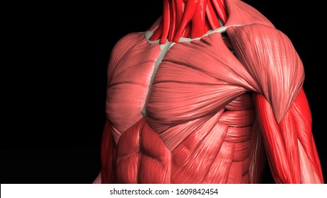
Male Chest Abdominal Muscles Anatomy In Blue X Ray Outline Full Color 3d Computer Generated Illustration On Black Background Stock Photo Alamy from c8.alamy.com Chest muscle anatomy the pectoralis major muscles also known as the pecs are located on the front of the rib cage and form the major muscles of the chest. Learn about each of these muscles, their locations, functional anatomy and exercises for them. These include pectoralis major, pectoralis minor, serratus anterior, and subclavius. (1) the pectoralis major, and (2) the pectoralis minor. The pectoral region is located on the anterior chest wall. Anatomy chart courtesy of fcit. Not everyone, however, has the chance to achieve stunning results when working on chest muscles. The pectoralis major muscles (also known as the pecs) are located on the front of the rib cage, and form the major muscles of the chest.
It's considered to be one of the most effective and reliable methods of measuring muscle activity.
Use the mouse scroll wheel to move the images up and down alternatively use the tiny arrows on both side of the image to move the images. Pectoralis major, pectoralis minor, serratus anterior, and subclavius. Muscles the dominant muscle in the upper chest is the pectoralis major. This mri chest thorax axial cross sectional anatomy tool is absolutely free to use. It contains four muscles that exert a force on the upper limb: (1) the pectoralis major, and (2) the pectoralis minor. The pectoralis major, pectoralis minor, serratus anterior and subclavius. Computed tomography (ct) of the chest can detect pathology that may not show up on a conventional chest radiograph(1). Ventral trunk muscles (overview) the trunk (torso) is the central part of the body to which the head and the limbs are attached. This page provides an overview of the chest muscle group. Muscles of the chest and their functions you have two mighty muscles on both sides of your chest: Muscles the major muscle in the chest is the pectoralis major. Chest the chest anatomy includes the pectoralis major, pectoralis minor, and the serratus anterior.
Ventral trunk muscles (overview) the trunk (torso) is the central part of the body to which the head and the limbs are attached. You have two pectoralis majors or pecs, one on each side of your chest. Injury to the pectoralis major can cause shoulder pain and limit your ability to use your arm fully. Use the mouse scroll wheel to move the images up and down alternatively use the tiny arrows (>>) on both side of the image to move the images.>>) on both side of the image to move the images. This mri chest (thorax) axial cross sectional anatomy tool is absolutely free to use.
Chest Anatomy Illustration Images Stock Photos Vectors Shutterstock from image.shutterstock.com Muscles of the head by label It's considered to be one of the most effective and reliable methods of measuring muscle activity. Muscles the major muscle in the chest is the pectoralis major. The pectoralis major muscles (also known as the pecs) are located on the front of the rib cage, and form the major muscles of the chest. See chest muscles anatomy stock video clips. Computed tomography (ct) of the chest can detect pathology that may not show up on a conventional chest radiograph(1). The muscles of the chest and upper back occupy the thoracic region of the body inferior to the neck and superior to the abdominal region and include the muscles of the shoulders. Muscles the dominant muscle in the upper chest is the pectoralis major.
Anatomy chart courtesy of fcit the pecs attach to the humerus near the shoulder joint and originate on the breastbone in the center of the chest.
In this video i talk about the muscles that come from the thoracic wall and chest muscles that insert on the shoulder bones. Injury to the pectoralis major can cause shoulder pain and limit your ability to use your arm fully. The torso muscles attach to the skeletal core of the trunk, and depending on their location are divided into two large groups: These important muscles control many motions that involve moving the arms and head — such as throwing a ball, looking up at the sky, and raising your hand. Ventral trunk muscles (overview) the trunk (torso) is the central part of the body to which the head and the limbs are attached. These include pectoralis major, pectoralis minor, serratus anterior, and subclavius. Human muscles · august 15, 2020. Related posts of chest muscles diagram muscle anatomy trivia. The pecs attach to the humerus near the shoulder joint and originate on the breastbone in the center of the chest. Anatomy chart courtesy of fcit. Pectoralis major, pectoralis minor, serratus anterior, and subclavius. Chest muscle anatomy the pectoralis major muscles (also known as the pecs) are located on the front of the rib cage, and form the major muscles of the chest. The chest or thorax is the region between the neck and diaphragm that encloses organs, such as the heart, lungs, esophagus, trachea, and thoracic diaphragm.
The muscles of the chest and upper back occupy the thoracic region of the body inferior to the neck and superior to the abdominal region and include the muscles of the shoulders. Muscles of the head by label These include pectoralis major, pectoralis minor, serratus anterior, and subclavius. These muscles help pull your arm across the front of your body. The fibers of the pectoralis muscles run like a fan across the.
Anatomy Study Chest Muscles By Dipnusurf On Deviantart from images-wixmp-ed30a86b8c4ca887773594c2.wixmp.com The muscles of the chest and upper back occupy the thoracic region of the body inferior to the neck and superior to the abdominal region and include the muscles of the shoulders. Muscles of the chest and their functions you have two mighty muscles on both sides of your chest: To know whether or not an exercise targets the right muscles or not, scientists use a type of test called electromyography (emg). Related posts of chest muscles diagram muscle anatomy trivia. See human chest anatomy stock video clips. These large muscles help you move your shoulder. Start with a pair of dumbbells extended above your chest. The beginner as well as advanced players meet with the problem when building a powerful chest.
Pectoralis major, pectoralis minor, serratus anterior, and subclavius.
Anatomy chart courtesy of fcit the pecs attach to the humerus near the shoulder joint and originate on the breastbone in the center of the chest. Here, we break down the anatomy of your chest muscles. Use the mouse scroll wheel to move the images up and down alternatively use the tiny arrows (>>) on both side of the image to move the images.>>) on both side of the image to move the images. The torso muscles attach to the skeletal core of the trunk, and depending on their location are divided into two large groups: Muscles of the head by label See human chest anatomy stock video clips. All about the chest muscles the chest anatomy includes the pectoralis major, pectoralis minor and the serratus anterior. The pectoral region is located on the anterior chest wall. The muscles of the chest and upper back occupy the thoracic region of the body inferior to the neck and superior to the abdominal region and include the muscles of the shoulders. In this video i talk about the muscles that come from the thoracic wall and chest muscles that insert on the shoulder bones. Human muscles · august 15, 2020. Muscles of the chest and their functions you have two mighty muscles on both sides of your chest: The muscles of the chest and upper back occupy the thoracic region of the body inferior to the neck and superior to the abdominal region and include the muscles of the shoulders.